

Discover the throughput and precision that is uniquely designed for translational researchers.
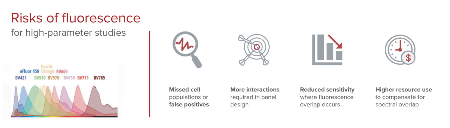
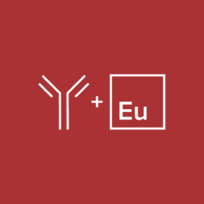
Imaging Mass Cytometry technology generates high-dimensional spatial data at subcellular resolution. IMC technology uses cytometry by time-of-flight (on which CyTOF™ systems are based) to overcome the multiplexing limitations of traditional immunohistochemistry (IHC) and immunofluorescence. By applying metal-tagged antibodies instead of fluorochromes, IMC technology has the unique capability to simultaneously stain, acquire and analyze 40-plus markers of interest on a tissue section without interference from autofluorescent tissues or management of spectral overlap.
IMC is the only technology with
No autofluorescence interference to image any tissue type
40-plus markers imaged simultaneously to get results faster
Integrated cell segmentation for faster interpretation
Batch staining of all slides for high-volume studies
Dual imaging and flow cytometry mode to maximize investment
IMC shows the true biology
High-plex imaging for all tissue types ― including lung, bone marrow, colon and brain ― without autofluorescence interference.
- Well-defined red signals from CD68
- Cellular structure is sharply defined by yellow pan-cytokeratin stain
- CD68 indistinct or missing
- Cellular structure diffuse

Breaking ground: Taking spatial biology into the clinic
Hear how the team at Navignostics is translating spatial insights into the clinic by leveraging spatial proteomics into reporting within 72 hours of sample receipt, revolutionizing treatment decisions for each cancer patient.
Jana Fischer, PhD | Co-Founder and CEO, Navignostics
Workflow
Get results faster
Hyperion™ Imaging Systems use a one-step staining and detection approach that enables samples to be simultaneously stained, acquired and analyzed.
Modularized panels
Swap markers without panel revalidation.

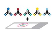
Simultaneous staining
Stain 40-plus markers for all slides at once.

One-step detection
Simultaneous imaging of 40-plus markers, including protein and RNA.

Precise signals
Image any tissue without autofluorescence.

Real-time analysis
Visualize 40-plus markers in 30 minutes.
A one-step staining and detection workflow
Imaging Mass Cytometry technology enables 40-plus markers that can be simultaneously stained, acquired and visualized. Other fluorescence-based approaches involve iterative rounds of staining, imaging and removal of fluorescent signals. The IMC workflow is without sequential immunostaining approaches, multiple slide treatment rounds or acquisition steps.
Need to ship or store slides?
A workflow ideal for high-volume samples,
clinical research trials and multi-site studies
- All-at-once batch staining of all slides to reduce technical variation
- Acquire at any time from shipped and/or stored slides that have been stained
- Analyze previously banked tissue slides to be correlated to known clinical outcomes
Applications


Key publications showcasing IMC technology