
See the next generation in spatial biology
High-throughput multiplexed whole slide tissue imaging: The Hyperion XTi Imaging System now offers the ability to automate slide loading for continuous acquisition and new whole slide imaging modes for rapid and detailed analysis.
Visualize tissue heterogeneity in 20 minutes.
One instrument, dual functions
Leverage the unique integrated dual modality of single-cell and spatial proteomic capabilities with the Hyperion XTi Imaging System and CyTOF XT systems. Take a deep dive into translational and clinical applications that explore new views of cellular composition within the tissue microenvironment. Watch this brief overview.
TECHNOLOGY
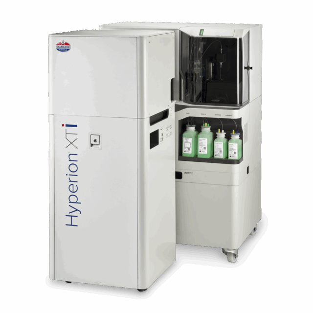
Powered by proven Imaging Mass Cytometry™ (IMC™) technology, the Hyperion XTi Imaging System is a multiplexed tissue imager that uses metal-tagged antibodies, instead of fluorophores, to simultaneously and reliably acquire 40-plus protein and RNA markers, without autofluorescence interference. IMC technology is the trusted choice for researchers and continues to be featured in high-impact publications – making it the most reliable tool for highly multiplexed imaging.
SEE DATA QUICKLY
Researchers can visualize 40-plus markers in real time, without managing time-consuming acquisition cycles – expediting the time to deeper analysis.
View the Hyperion XTi image lookbook to learn how whole slide and cell imaging modes can be applied.
Represents Hyperion XTi Preview Mode for a large tissue section over 25 minutes. Researchers can select and view from any markers on the panel during acquisition.
SEE DATA CLEARLY.
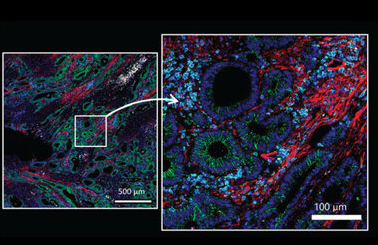
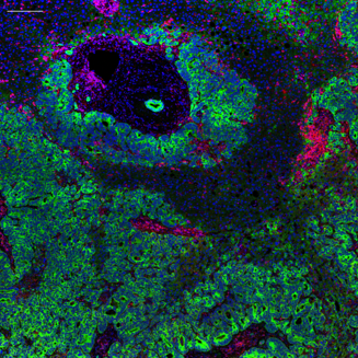
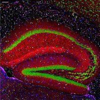
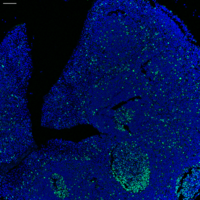
Clearly define areas in any tissue type ― including lung, bone marrow, colon and brain ― without background autofluorescence and spectral overlap.
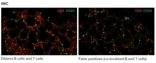
In bone marrow, cyclic immunofluorescence data (right) shows false positives, highlighted by co-localization of B cells (CD20+) and T cells (CD3+) (yellow). Conversely, distinct B cells (green) and T cells (red) can be seen with IMC technology (left).
Discover the power of Imaging Mass Cytometry technology

Entering an era of new biology with rapid, high-throughput imaging
“Faster means more samples, more data. We can push through more of these small samples or we can do whole slides on the Hyperion XTi”
Hartland Jackson, PhD
Investigator
Lunenfeld-Tanenbaum Research Institute, Sinai Health
WORKFLOW
40 slides. 40-plus markers. 24 hours.
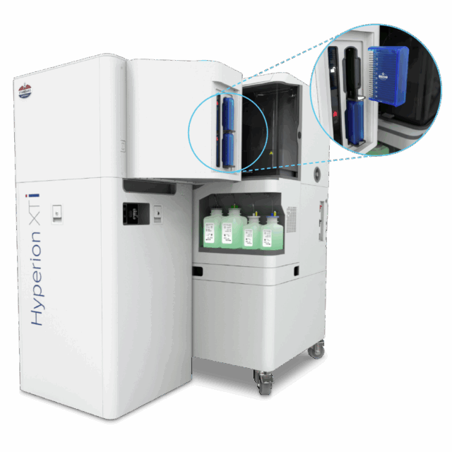
Load up to 40 slides and walk away.
Incorporate a new level of throughput and efficiency with the Hyperion XTi Slide Loader. The optional slide loader enables automatic loading and acquisition of up to 40 slides. It is equipped with a barcoded slide reader for easy sample identification, providing researchers with peace of mind when managing precious tissue samples.
See more from your samples.
Do more with your time.
Flexible Imaging Modes for Different Needs
Preview Mode
Number of markers: 42
Acquisition time: 20 minutes
Sample: colon cancer (25 mm X 15 mm)
Cell Mode
Number of markers: 42
Acquisition time: 2 hours
Sample: colon cancer (2 mm X 2 mm)
Resolution: 1 Um
Tissue Mode
Number of markers: 42
Acquisition time: 5 hours and 50 minutes
Sample: Breast cancer (24 mm X 16 mm)
Resolution: 5 Um
Multiplexed imaging – without compromising speed
An end-to-end workflow to get results faster
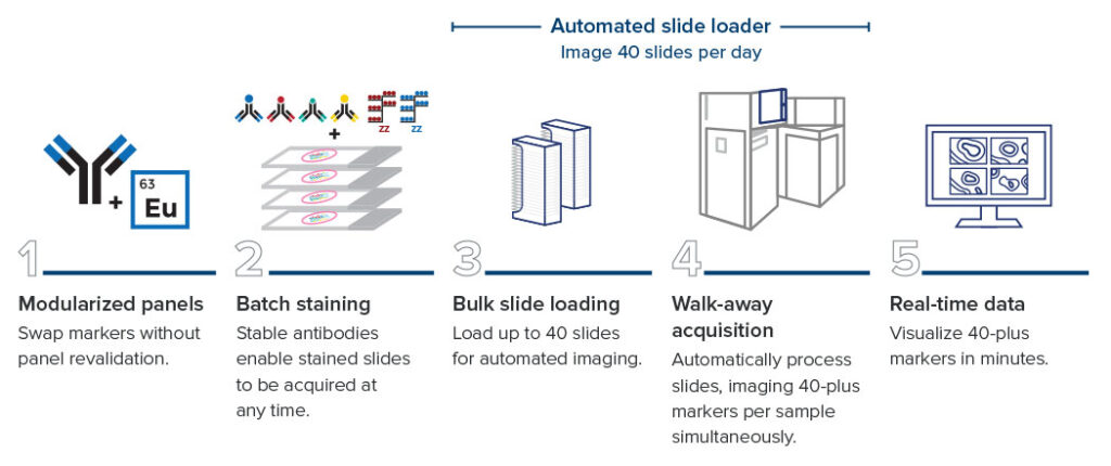
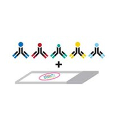
A one-step staining and detection workflow
Imaging Mass Cytometry technology enables 40-plus markers that can be simultaneously stained, acquired and visualized. Other fluorescence-based approaches involve iterative rounds of staining, imaging and removal of fluorescent signals. The IMC workflow is without sequential immunostaining approaches, multiple slide treatment rounds or acquisition steps.
Same-slide omics
Capture 40-plus markers of spatial proteomic data after RNA detection using the same slide. The Hyperion XTi workflow provides the ability to add essential complementary proteomic insights to previously acquired spatial transcriptomic data, with an option to also add in H&E staining, if desired.
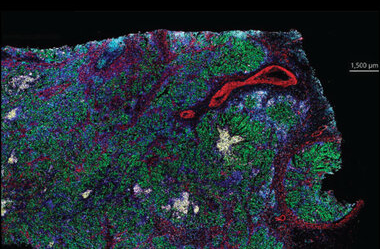
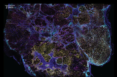
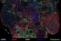
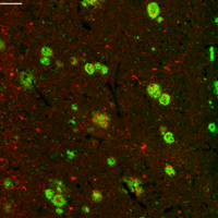
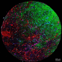
This image highlights the clarity of IMC to detect both low and high abundance proteins in human hepatocellular carcinoma using the Hyperion XTi Imaging System and a 43-marker panel combined from two ready-to-go base and expansion panels.
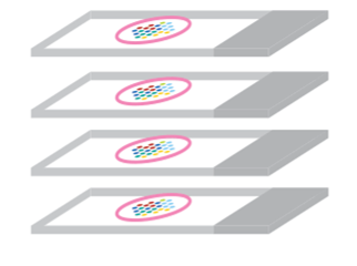
Need to ship or store slides?
- All-at-once batch staining of all slides to reduce technical variation
- Acquire at any time from shipped and/or stored slides that have been stained
- Analyze previously banked tissue slides to be correlated to known clinical outcomes
Let us do the work for you.
Did you know Standard BioTools has an in-house service lab? Send us your samples and we will send you the data.
Applications
IMAGING PANELS
1. Start with ready-to-go human and mouse panels.
2. Easily add the Maxpar™ IMC Cell Segmentation Kit to your panel to solve the most important step in spatial biology.
3. Want to further customize your panel? Easily swap targets of interest from an ever-growing catalog of Maxpar IMC antibodies and panels.
Resources
CUSTOMER STORIES
Read success stories from our customers.


CyTOF Software v9.3
CyTOF™ Software v9.3 offers instrument control and data acquisition functionality for both imaging and flow applications with the Hyperion™ XTi Imaging System, CyTOF XT System and CyTOF XT PRO Syst... v9.3 September 01, 2025
MCD Viewer
MCD™ Viewer is post-acquisition data processing software that allows users to visualize, review and export Imaging Mass Cytometry™ data acquired with the Hyperion™ Imaging System and CyTOF™&nb... v1.0.560.6 August 12, 2021
MCD Smartviewer V1.1.0
MCD™ SmartViewer v1.1.0 is post-acquisition data processing software for visualization, clustering and spatial analysis of data acquired in Tissue Mode using the Hyperion™ XTi Imagin...
Visiopharm Software
Phenoplex is a complete workflow software for multiplex tissue images in Spatial Biology, built on Visiopharm’s best-in-class AI, with interactive verification steps throughout. November 17, 2025
ILASTIK
ilastik is an open source software for image analysis and machine learning. Using machine learning, ilastik software trains custom workflows for accurate cell segmentation and classification. v1.4.0 February 20, 2023
HISTOCAT
The histoCAT software is an innovative computational Imaging Mass Cytometry™ analysis toolbox that enables comprehensive analysis of cellular phenotypes and their interrelationships within the... v1.761 June 16, 2020
GET MORE INFORMATION
Interested in Standard BioTools™ Imaging Mass Cytometry products or need a quote? Contact our sales team for product orders, quotes or other inquiries.