Cell Segmentation
High-plex image analysis starts with robust cell segmentation
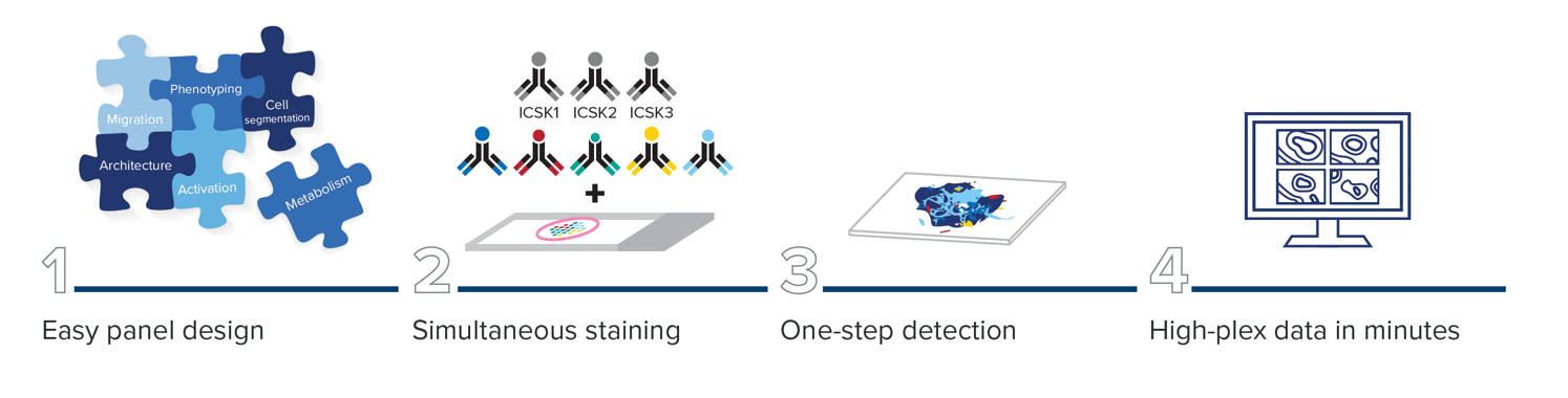
The advent of high-plex imaging techniques demands more accurate and reliable cell segmentation. To interpret the significant amount of data that comes from high-plex single-cell imaging approaches such as Imaging Mass Cytometry™ (IMC™) technology, effective assignment of cellular borders is a key analytical challenge. The Maxpar™ IMC Cell Segmentation Kit facilitates an end-to-end workflow for single-cell data analytics and is an important first step to getting improved high-plex cell segmentation regardless of which pipeline is used.
Segment with confidence using the Maxpar IMC Cell Segmentation Kit.
The Maxpar IMC Cell Segmentation Kit enables enhanced nucleus and plasma membrane demarcation. This integrated solution easily solves the most critical step in high-plex spatial imaging without limiting your 40-plus-marker panel.
Learn more about the Maxpar IMC Cell Segmentation Kit.
Facilitating an end-to-end workflow with the Maxpar IMC Cell Segmentation Kit
Spatial biology is complex.
Experiments don’t have to be.
Unfamiliar with the specifics of cell segmentation?
Our October 2021 IMC Summit: Uncovering Spatial Biology had a data analysis session dedicated to understanding the importance of cell segmentation in the data analysis pipeline. Watch the recording of this session, which has two presentations, one by Hartland Jackson, PhD, and a joint talk by Jay Hirota, PhD, and Kjetil Ask, PhD.
Customer Stories
Learn more about how our customers are leveraging Standard BioTools™ technology
Febe van Maldegem, PhD, and Karishma Valand

Imaging the dramatic remodeling of the lung tumor microenvironment
Corinne Ramos, PhD, MBA

Advancing drug development with molecular imaging
Jana Fischer, PhD

Paving a new road for spatial biology in precision medicine
Yongpan Yan, PhD

An approach that combines molecular biology, immunology and cell biology tools to uniquely
Melissa Davis, PhD

Defining spatial characteristics using Imaging Mass Cytometry™ technology
Rebecca Ihrie, PhD

How CyTOF™ technology enabled a look into the heterogeneity of glioblastoma.
Xianting Ding, PhD

High-dimensional single-cell analysis enhances precision medicine
Dr. Dexi Chen, PhD, MD

Achieving multi-omic insights with microfluidics and CyTOF™ technology
Denis Schapiro, PhD

The interactive histoCAT toolbox unifies the cytometry and imaging communities
Bernd Bodenmiller, PhD

Bernd Bodenmiller on revealing new insights into tumors with Imaging Mass Cytometry™.
David L. Rimm, MD, PhD

David Rimm on the value of Imaging Mass Cytometry™ for immunology applications at Yale
Akil Merchant, MD

Hear how researchers are using Imaging Mass Cytometry™ technology.
Hartland Jackson, PhD

Entering an era of new biology
Daniel Schulz, PhD

Discovering a chemokine’s purpose in fighting tumors using RNA and protein co-detection
RELATED BLOG POSTS
-
Have autofluorescence? Now you don’t.
Learn how Imaging Mass Cytometry platforms permit cleaner detection of surface and intracellular targets for quantifiable phenotyping and cell function assessment
For Research Use Only. Not for use in diagnostic procedures. Patent and License Information: www.standardbio.com/legal/notices. Trademarks: www.standardbio.com/legal/trademarks. Any other trademarks are the sole property of their respective owners. ©2025 Standard BioTools Inc. All rights reserved.