Standard BioTools at AACR 2023: Advancing the Frontiers of Cancer Science and Medicine
The American Association for Cancer Research will be holding its annual meeting from April 14–19.
We are proud to serve a large and growing community of researchers who are making breakthroughs in translational and clinical cancer research. We appreciate your trust in Standard BioTools™ to help capture critical immune data from sample collection to novel insights with confidence ― so you don’t miss the unexpected … or even what you expect to find.
Visit Booth 1224 to discuss how you can improve your productivity and get reliable results faster. Plus, come see what we’ve got in store!

Learn and network
We are thrilled to invite you to an exciting event to socialize and network with leaders in the cancer research community.
Connect with colleagues and potential collaborators who are using high-parameter flow cytometry and Imaging Mass Cytometry™ to shape the future of clinical immunotherapies. Hear about new tools and techniques that can make a difference in how you identify cellular dynamics and spatial features.
Advancing Multiplex Spatial Analysis to New Limits
→ Speaker: Hartland Jackson, PhD
Investigator, Lunenfeld-Tanenbaum Research Institute
→ Date and time: Monday, April 17
7:00 pm | Cocktails and networking
8:00 pm | Dinner
→ Venue: The Hampton Social*
9101 International Drive, Orlando, FL 32819
* Space is limited. Please RSVP to secure your spot.
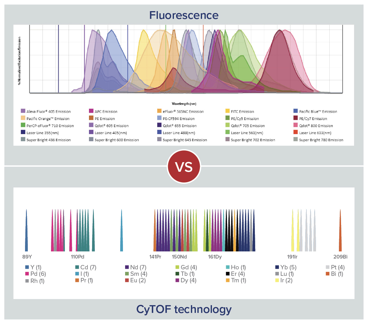
CyTOF technology
Featured in over 2,000 publications and 200 clinical trials, CyTOF® technology provides reproducible and standardized workflows, simple and easy-to-modify panel design and precise measurement of all markers. This high level of data quality empowers immune profiling for any phase of clinical research.
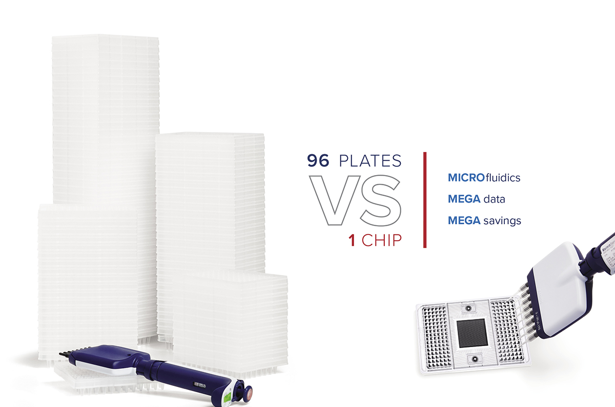
Microfluidics technology
Integrated into a broad range of applications, microfluidics technology accelerates genomic analysis by enabling 9,216 reactions in a single run, requiring less time, less sample and less reagent than conventional protocols. Now you can perform real-time PCR applications and NGS library prep on a single system.
SCIENTIFIC POSTERS
Optimized Human Immunophenotyping Panels Enhance the Flexibility for High-Dimensional Flow Cytometry Analysis with CyTOF
Simplifying High-Parameter Phenotypic and Functional Characterization of Immune Cells
50-Parameter Flow Cytometry by CyTOF Empowers Comprehensive Single-Cell Immune Profiling of Pulmonary Immunosenescence in Aged Mice
High-Plex Co-Detection of RNA and Protein to Explore Tumor-Immune Interactions Utilizing RNAscope™ with Imaging Mass Cytometry
Identifying Pathophysiological Features of Mouse Tumors Using Imaging Mass Cytometry
Imaging Mass Cytometry Enables Identification of Distinct Tissue Phenotypes in Highly Autofluorescent Lung and Colon Cancer Tissues, Displaying Consistent Data Across Serial Sections
Immuno-Oncology Study to Profile the Tumor Microenvironment in Multiple Human Cancers Using High-Plex Imaging Mass Cytometry
Increasing Plexity of Imaging Mass Cytometry for Tumor Tissue Analysis
Neuro-Oncology Imaging Mass Cytometry Panels Enable Spatial Investigation of Brain Tumor Microenvironment
Learn more about the technology
For Research Use Only. Not for use in diagnostic procedures. Patent and License Information: www.standardbio.com/legal/notices. Trademarks: www.standardbio.com/legal/trademarks. Any other trademarks are the sole property of their respective owners. ©2025 Standard BioTools Inc. All rights reserved.