Flexible panel design, powerful results
Modular panels allow for an unprecedented look into cell populations

Accurately identifying different cell types within a heterogeneous population relies on good panel design, which, depending on the technology, can be time-consuming and complex. With fluorescence-based cytometry, the issues of spectral overlap and autofluorescence mean designing high-parameter experiments can be complicated. Overlapping emission spectrums can result in difficult-to-determine cellular relationships, while a finite number of fluorophore tags means panel builds are limited.
Enter CyTOF™ technology. Mass cytometry uses rare-earth metals instead of fluorophores, permitting easy panel design and modification and deep analysis of cell types without having to consider co-expressed markers, signal overlap or the brightness of individual fluorophores. CyTOF systems can analyze multiple markers at once, allowing researchers to look at everything within a sample without having to go back and rerun an experiment with a different panel. In addition, mass cytometry instruments have 135 channels, which means they can easily detect 40–60 markers, with the potential to increase that number.
From backbone panels to precise objectives
Immunophenotyping, a technique used to identify and classify cells based on the proteins they express, is used in research to help diagnose diseases, such as types of leukemia and lymphoma. By coupling antibodies with metal tags, oligos or fluorophores, it allows researchers to detect the presence, absence and expression patterns of proteins. The patterns are then used to phenotype or classify the cells. By increasing the numbers of parameters in a panel, researchers can customize their panels to meet their specific experiment needs and further phenotype their samples.
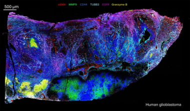
Modular pathologist-verified Imaging Mass Cytometry™ (IMC™) panels allow researchers to explore the relationship between cellular function and location within the tissue. IMC technology uses cytometry by time-of-flight (on which CyTOF systems are based), overcoming the multiplexing limitations of traditional immunohistochemistry and fluorophore-based technologies such as immunofluorescence. Pre-validated IMC panels overcome the common challenges experienced with fluorescence-based technologies to map function across tissues.
Standard BioTools™ modular IMC panels allow researchers to easily and quickly build panels with seamless compatibility, with the flexibility to add custom targets as needed. Our panel-building flyer outlines how ready-to-go pre-validated panels overcome the common challenges, complexity and labor involved with fluorescence techniques and IHC and allow researchers to see the whole picture.
Immuno-oncology focus
Quickly apply spatial context to immuno-oncology research by combining ready-made modular IMC panels that cover a breadth of immune profiling and functional targets. Researchers can start with the Human Immuno-Oncology IMC Panel, 31 Antibodies and add compatible panels to further characterize tumor and immune cell subtypes.

- Start with the Human Immuno-Oncology IMC Panel, 31 Antibodies (PN 201509). This panel enables evaluation of critical pathophysiological parameters of the human tissue microenvironment using 31-plex IMC technology.
- Add the Human Immune Cell Expansion IMC Panel, 7 Antibodies (PN 201516). This panel provides additional cell phenotyping markers for identification of lymphoid and myeloid cell subtypes of immune cell infiltrates in tumors.
- Perform cell segmentation with the Maxpar™ IMC Cell Segmentation Kit (PN 201500). The ICSK facilitates an end-to-end workflow for single-cell data analytics. The kit contains three individual plasma membrane markers conjugated to 195Pt, 196Pt and 198Pt, and is designed to improve plasma membrane demarcation in cell segmentation. The ICSK uses the markers in combination to label plasma membranes of all cell types homogeneously within the tissue. Each marker provides variable staining that is dependent on the cell and tissue type, ensuring coverage of all cells in a tissue.
- Cell-ID™ Intercalator-Ir (PN 201192A/ 201192B) can be combined with the ICSK for improved cell segmentation. Cell-ID Intercalator-Ir is a cationic nucleic acid intercalator that contains natural abundance iridium (191Ir and 193Ir). When cells are stained with Intercalator-Ir, it binds to cellular nucleic acid, and detection of both stable isotopes enables identification of nucleated cells in CyTOF analysis.
READ MORE: Check out our app note Reveal Heterogeneity of Tumors with Whole Slide Imaging, which details how to detect clinically relevant components of the TME to provide insight into the state of the tumor tissue and empower researchers to evaluate parameters related to patient survival and treatment.
Neurodegenerative focus
Leverage spatial cell phenotyping with IMC technology for high-quality neurological and immunological insights. Modular panels can help researchers analyze over 40 proteins simultaneously in neoplastic or neurodegenerative tissues without interference from the autofluorescence commonly observed in brain tissue.

- Start with the Human Basic Immune IMC Panel, 4 Antibodies (PN 201518) to phenotype key lymphoid and myeloid cell subtypes; the Human Lymphoid IMC Panel, 4 Antibodies (PN 201512) to delineate lymphoid cell subtypes of immune cell infiltrates in tumors; the Human Myeloid IMC Panel, 6 Antibodies (PN 201513) to delineate myeloid cell subtypes of immune cell infiltrates in tumors; and the Human Basic Tissue Architecture IMC Panel, 3 Antibodies (PN 201517) to identify the underlying cellular and structural architecture of normal and diseased human tissues.
- Add the Alzheimer’s Disease IMC Panel, 3 Antibodies (PN 9100007) to study Alzheimer’s disease and tauopathies or amyloidopathies; and the Maxpar Neuro Phenotyping IMC Panel Kit (PN 201337) to identify major cell types such as neurons, astrocytes, microglia, oligodendrocytes and endothelial cells and their functional state in the central nervous system.
- Perform cell segmentation with the Maxpar IMC Cell Segmentation Kit. Cell-ID Intercalator-Ir (PN 201192A/ 201192B) can be combined with the ICSK for improved cell segmentation.
READ MORE: Check out our app note Exploring Neurodegenerative Diseases with Imaging Mass Cytometry to learn more about how IMC technology captures key biological insights and provides a comprehensive multimodal approach to understanding the complex pathology of neurodegenerative conditions.
Mouse immuno-oncology and neurology focus
Use the Maxpar OnDemand Mouse Immuno-Oncology IMC Panel Kit to evaluate critical pathophysiological parameters of the mouse tumor microenvironment (TME). Researchers can combine the kit or its four modular subpanels with the Maxpar Neuro Phenotyping IMC Panel Kit for comprehensive brain tumor characterization and assessment of neuronal inflammation, activation and development.

- Start with the Maxpar OnDemand Mouse Immuno-Oncology IMC Panel Kit (PN 9100005) to enable the evaluation of critical pathophysiological parameters of the murine TME
- Add the Maxpar Neuro Phenotyping IMC Panel Kit
- Perform cell segmentation with the Maxpar IMC Cell Segmentation Kit. Cell-ID Intercalator-Ir (PN 201192A/ 201192B) can be combined with the ICSK for improved cell segmentation.
READ MORE: Check out our app note Unravel the Complexity of Mouse Brain Tumors to learn more about conducting detailed mouse neuro-oncology studies that can uncover crucial insights into tumor development, progression and treatment.
Specific panels for specific research
Standard BioTools IMC panels make study design simple. By tailoring marker selection to specific biological pathways or research questions, researchers can uncover tumor-immune interactions, map localized functional responses and characterize cell features, revealing spatial biomarkers essential for understanding disease progression and immune response – all in a single experiment.
