Have autofluorescence? Now you don’t.
Learn how Imaging Mass Cytometry permits cleaner detection of surface and intracellular targets for quantifiable phenotyping and cell function assessment

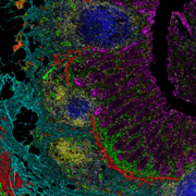
Autofluorescence, the natural fluorescence that occurs in biological structures, can hamper your work in the lab. Importantly, it can affect the sensitivity of assays by leading to poor-quality images that make it hard to detect stains targeting specific cells. Depending on tissue type and signal intensity, autofluorescence can result in a false positive population or an artificial increase in the percentage of positive cells, especially if you’re working with highly autofluorescent tissues like lung, gut, brain or skin. When the background interferes, you may be unable to distinguish cell populations or complex architecture, which presents a significant challenge.
But grappling with autofluorescence isn’t a forgone conclusion. Because Imaging Mass Cytometry™ uses rare-earth metals as labels for antibodies instead of fluorophores, the background is non-existent, as these metals are not present in tissue samples. Additionally, since detection peaks are distinct and do not overlap, there is very little background overall. This permits cleaner detection of surface and intracellular targets for quantifiable phenotyping and cell function assessment.
IMC™ produces high-quality data similar to other imaging techniques, such as fluorescent multiplex imaging. However, fluorescent imaging technologies are prone to an abundance of autofluorescent artifacts, while these problems are virtually nonexistent in IMC. Because other fluorescence-based approaches have a vastly lower dynamic range than IMC, heterogeneous systems with big differences between the lowest- and highest-expressed molecules often appear overexposed, with signals showing up in unwanted cells.
Analyzing fluorescence data can also take time and energy and can even result in a loss of experiment sensitivity. The process of gating—isolating populations using molecules that are visualized by fluorescence in a unique emission spectrum—is complex, often involving multiple regions of interest or requiring backgating if you’re unsure of your process. In addition, tighter gates may mean you’re missing a crucial population.
Using IMC means you don’t have to deal with all of that. With IMC, you can effectively assign cellular borders, resulting in accurate and reliable cell segmentation.
More on why IMC reduces autofluorescence
Check out other blog posts
Familiar with IHC?
Then you already know IMC.
If IHC is your comfort level, IMC lets you maintain that confidence in your research while moving it forward for insights you didn’t see coming.
Invest in Automation to Save Money in the Long Run

Where should managers put their focus to ensure their labs run smoothly? Learn about a new metric that can help improve lab efficiencies and cost savings for years to come.
To automate or not to automate?

Considerations for when to automate, from common drivers to what you really should be thinking about.
Unless explicitly and expressly stated otherwise, all products are provided for Research Use Only, not for use in diagnostic procedures. Find more information here.