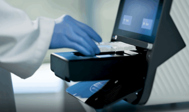
Products Technologies Imaging Mass Cytometry IMC Trending Topics and Publications IMC Trending Topics News

IMC Trending Topics News

background pattern

IMC Trending Topics News Archive

Check this page to see all news and stories about researchers who are using IMC to make an impact.

  • Congratulations to the five Canadian cancer research teams that won the 2022 Terry Fox New Frontiers Program Project Grant competition! The teams will be able to expand their research into key areas of cancer treatment.
    One of the longstanding award holders, a team led by Dr. Christian Steidl, head of lymphoid cancer research at BC Cancer, continues its groundbreaking research using novel technologies, including single-cell sequencing and Imaging Mass Cytometry™, to describe changes in tumors, cell by cell, and visualize the altered micro-architecture of lymph nodes.
    “I am optimistic in saying with the depth and breadth of information we can derive from these samples, we can build complete models of how lymphoma evolves, and we can translate these results into novel therapies to obtain higher cure rates and fewer side effects for patients,” Steidl said in a press release.

  • At the American Society of Nephrology Kidney Week in November, Lloyd Cantley, MD, Professor, Vice Chair of Research and Co-Director of Education, Yale Center for Clinical Investigation, presented Spatial Analysis of the Kidney: Imaging Mass Cytometry of the Human Kidney. The meeting brings together kidney professionals from around the globe to exchange new research findings, learn about the latest clinical and scientific advances in the field and engage in exciting discussion with leading international experts.
  • The Bodenmiller Lab released the newest versions of their imcdatasets, cytomapper and imcRtools RStats packages on Bioconductor. These packages form a software suite to analyze and visualize highly multiplexed imaging data. The imcdatasets package provides direct access to publicly available Imaging Mass Cytometry datasets. The cytomapper package allows the visualization of multichannel image composites and maps single-cell information to segmentation masks. The imcRtools package provides spatial single-cell analysis approaches to characterize tissue structures and cell interactions.

  • The Black in Cancer Conference was held in collaboration with Cancer Research UK, showcasing new findings from cancer researchers around the world and including tools to model single-cell biology data from technologies like Imaging Mass Cytometry.

  • An Imaging Mass Cytometry workshop kicked off Spatial Biology US, a meeting for those integrating spatial biology and multi-omics approaches into their research. Presenters discussed the latest advancements in applying spatial data to garner deeper insights and overcoming key analysis challenges.

  • Thank you to Thomas Ashhurst, PhD, for presenting his work at this month’s CYTO® 2022 workshop – Integration and Analysis of High-Dimensional Single-Cell Cytometry and Imaging Data in a Murine Model of Viral Encephalitis.
  • Adriano Luca Martinelli and Maria Anna Rapsomaniki at IBM Research Europe in Zurich introduced a new computational framework, ATHENA, that facilitates the visualization, processing and analysis of tumor heterogeneity from spatial omics measurements, including from Imaging Mass Cytometry.

  • Melissa B. Davis participated in a National Breast Cancer Coalition panel discussion on healthcare disparities in the US and how to facilitate change in the movement toward health equity for all. She was also interviewed for the broadcast news story, “Racial and ethnic disparities in breast cancer outcomes,” which appeared on NY1 News on May 4.
  • Thanks to Anne Bärenwaldt at University of Bern and Inselspital who collaborated with us for a great Helios™ and Hyperion Imaging System kickoff in May! What a great Imaging Mass Cytometry community in Switzerland.
  • Congratulations to Tobias Boettler and team at the University of Freiburg for their April publication exploring the pathophysiology of known cases of liver inflammation following SARS-CoV-2 infection or vaccination. The study used Imaging Mass Cytometry for spatial immune profiling of liver biopsy tissue.
  • Thank you to the Abu Dhabi Stem Cells Center for their time training with the team of Antonio Bencomo-Hernandez on the Hyperion Imaging System. We can't wait to see the results from this amazing team on stem cell therapy research!
  • Kudos to the Newcastle University Flow Cytometry Core Facility, which supported a new publication by Jack Leslie, Derek Mann and team with some nice data on the Hyperion Imaging System describing how manipulation of neutrophils assists immunotherapy for liver cancer.

  • Congratulations to Mathieu Lajoie at the Goodman Cancer Institute, McGill University, whose spatial graph image is on the April 2022 Science Immunology cover! Mathieu is an author on the recent paper, Spatially mapping the immune landscape of melanoma using Imaging Mass Cytometry, in which the team used IMC to characterize the immune microenvironment of melanoma patient samples, allowing for the identification of prognostic biomarkers for immunotherapy.
  • ImaBiotech will present a new "Immune Signature Panel" as part of its Imaging Mass Cytometry Services at the AACR22 meeting in New Orleans. Read the news!

  • Here’s a great read about the IMMUcan project and how researchers are working toward highly personalized tumor profiling for cancer patients. Daniel Schulz, PhD, and the project team use IMC to generate images from small tissue sections and have developed an approach to zoom in on select regions that capture the immune cell heterogeneity.

  • In early February, the winners of the 2022 Terry Fox New Investigator Award were announced. One of the winners is Hartland Jackson, PhD, an investigator at the Lunenfeld-Tanenbaum Research Institute at Sinai Health. With this grant, Jackson’s team will use IMC, digital pathology and systems biology to better understand how the Hippo pathway controls immune response to cancer.

  • Researchers from the King’s College London Schools of Cancer and Pharmaceutical Sciences and Immunology and Microbial Sciences and the Cancer Systems Biology laboratory at the Francis Crick Institute, led by Francesca Ciccarelli, PhD, have developed a new software for the analysis of data from multiplexed imaging technologies such as IMC. “When we started to work with high-dimensional IMC data a couple of years ago, there were not many tools for their analysis,” Ciccarelli says. “It has been a lot of fun to develop our own tool thanks to the talent of Michele Bortolomeazzi, a PhD student” funded by the Cancer Research UK King's Health Partners Centre.

  • A team at Children’s Hospital Los Angeles is turning to IMC to assist in its search for acute liver failure biomarkers in children. Johanna Ascher Bartlett, MD, a pediatric gastroenterology fellow, received a Broad Clinical Research Fellowship Award from USC to support her study. The team is examining 25 different immune cell markers in an effort to identify a predictive pattern that could indicate patient outcome and potentially save children from undergoing transplants and immunosuppression.


Unless explicitly and expressly stated otherwise, all products are provided for Research Use Only, not for use in diagnostic procedures. Find more information here.