
IMC Trending Topics and Publications
You want to get the most out of your precious samples, to answer complex questions through deep interrogation and analysis of relevant data, to understand the role of spatial biology in health and disease and to publish and showcase your research results.
The Power of IMC
Learn how Imaging Mass Cytometry™ (IMC™) technology is used as a vital spatial biology tool.
IMC Trending Topics
IMC Trending Topics articles, toolbox, news and FAS Favorite feature the great work performed by IMC users.
IMC Publications and Posters
Find current IMC bibliographies and posters, read the new FAS Favorite in publications and view charts that demonstrate the published use of IMC.
The power of IMC
- Subcellular resolution: Allows accurate and quantifiable phenotyping and cell function assessment
- No autofluorescence: Reliable results when working with highly autofluorescent tissues like lung, gut, brain and skin
- No time-consuming cyclic protocols: High-plex protein detection from FFPE or frozen tissue sections
- Proven: Hundreds of hands-on users and more than 130 peer-review published references

IMC Trending Topics articles
These articles cover the latest research and publications involving IMC technology, exploring topics such as immune landscape investigation, biomarker discovery and methods associated with therapeutics development.
Immuno-oncology TRENDING topics ARTICLE |
||
|
|
Immuno-oncology is a burgeoning field with many promising approaches to improving a patient’s outcome through immunotherapy. IMC technology has been influential in understanding underlying mechanisms of immune response and in developing strategies for new treatments. |
|
IMC Trending Topics toolbox
Recommended resources grouped to provide you with knowledge, ideas and tips on selected topics.

Robust cell segmentation is essential for high-plex image analysis. The resources below address key cell segmentation challenges. They also help if you are unfamiliar with the general why and how of cell segmentation. Included is information about the Maxpar™ IMC Cell Segmentation Kit, which facilitates an end-to-end workflow for single-cell data analytics and is an important first step to improve high-plex cell segmentation regardless of which pipeline is used.
- Information: Get more information from the Maxpar IMC Cell Segmentation Kit Information Sheet or the FAQ document.
- Related blog post: A groundbreaking new partnership
- Related blog post: Four experts share their image analysis tools and key insights

Staining for tissue imaging on the Hyperion™ Imaging System is similar to standard immunohistochemistry techniques in terms of tissue collection, tissue processing and the staining itself. Optimized staining is important in order to carry out extensive characterization of the tissue microenvironment. There are numerous examples of panels of up to 40 markers described for FFPE human tumor sections, and a protocol for frozen tissue sections is also available.
- Protocol: Imaging Mass Cytometry Staining User Guide
- Publication: A 40-marker panel for high dimensional characterization of cancer immune microenvironments by Imaging Mass Cytometry. (Ijsselsteijn et al.)
- Publication: High dimensional Imaging Mass Cytometry panel to visualize the tumor immune microenvironment contexture. (Elaldi et al.)
- Webinar: In a webinar sponsored by Cell Signaling Technology, Noel de Miranda, Principal Investigator of the immunogenomic group at Leiden University Medical Center, discusses critical considerations for optimizing Imaging Mass Cytometry technology.
Featured NEWS
IMC Trending Topics news
Check this feature regularly to see a current selection of exciting news and stories about researchers who are using IMC technology to make an impact.
DECEMBER 2023
- A summary of Distinguishing keratoacanthoma from well-differentiated cutaneous squamous cell carcinoma using single-cell spatial pathology by Veenstra et al. published in the December 2023 issue of the Journal of Investigative Dermatology highlights the use of RNA sequencing and Imaging Mass Cytometry technology to find features that differentiate keratoacanthoma and cutaneous squamous cell cancer.
Read the article -
Congratulations to Akil Merchant and team at Cedars-Sinai Cancer on their recent publication in the Journal of Clinical Oncology. The work outlines a new way to predict whether lymphoma will recur in patients treated with a bone marrow transplant and is one of the first to use spatial profiling to predict patient outcomes that could lead to more precisely targeted treatment.
“This work opens new avenues for the development of spatial biomarkers that can guide treatment of cancer patients in a precise, targeted way,” said Dan Theodorescu, MD, PhD, Director of Cedars-Sinai Cancer and the PHASE ONE Distinguished Chair. “Translation of this leading-edge research to a test that can be clinically deployed will help bring the promise of precision medicine to increasing numbers of patients – and will eventually benefit patients with many cancer types.”
Read the news
NOVEMBER 2023
- A press release from Oncolytics Biotech describes how novel IMC technology enables a closer look at the TME post-pelareorep/atezolizumab/letrozole treatment and affirms pelareorep’s ability to increase PD-L1+ cells and T cell infiltration in the tumor microenvironment (TME).
The positive translational data, presented at the Society for Immunotherapy of Cancer’s (SITC) 38th Annual Meeting, completes the AWARE-1 breast cancer window-of-opportunity study and supports conduct of a registrational program for pelareorep.
Read the article
OCTOBER 2023
- Laura Kuett recently completed her PhD at the University of Zurich and talked to SLAS Europe 2023 about her work using 3D Imaging Mass Cytometry technology to advance breast cancer research.
Watch the video
Read the interview - In an October webinar presented through Genetic Engineering & Biotechnology News, Sammy Ferri-Borgogno, an Instructor in the Department of Gynecologic Oncology and Reproductive Medicine at MD Anderson Cancer Center, discussed how a multi-omic approach to tissue imaging can reliably characterize the TME in ovarian cancer.
Watch the video
FAS Favorite
Check in regularly to see a featured publication recommendation from one of our expert Field Applications Scientist (FAS) team members.

MAY | JUNE
Wendell Smith, Senior Field Applications Scientist: Proteomics, recommends a Frontiers in Immunology publication by N. Ung et al., Adaptation of Imaging Mass Cytometry to explore the single cell alloimmune landscape of liver transplant rejection.
Principal Investigator: Juliet Emamaullee, MD, PhD, FRCSC, FACS, attending physician and assistant professor of clinical surgery from the surgery department at Keck School of Medicine of USC.
 Why Wendell recommends this publication: “I like this paper because it highlights our technology in a new area for Standard BioTools concerning our IMC platform: organ transplant rejection. This is an area of extreme importance both clinically and scientifically. The publication specifically utilizes our IMC technology to investigate the alloimmune microenvironment at a single-cell resolution during clinical rejection episodes.
What is also interesting about this paper is it was the first publication that I am aware of that utilized a set IMC panel that I helped design for the Children’s Hospital Los Angeles (CHLA) imaging core. The authors used this panel to characterize the immune landscape of chronic rejection (CR) in clinical tissue samples obtained from liver transplant recipients. This demonstrates how well a validated panel can work if cores choose to establish an appropriately validated panel.”
IMC publications
Join the hundreds of researchers gaining deep spatial insights using Imaging Mass Cytometry technology.
Get the complete IMC bibliography now, with over 130 peer-reviewed publications.
See more than 130 Publications
More than 50 publications highlight the use of high-plex imaging by IMC technology to investigate the spatial biology of the complex TME and its role in furthering cancer and immuno-oncology research.

IMC Posters
Learn more technical details about IMC technology by viewing some of our recently presented posters.
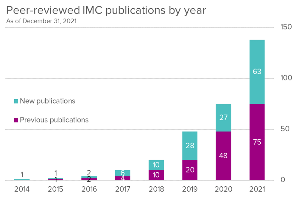
As of December 31, 2021, there are 138 total peer-reviewed publications for IMC, with 63 from 2021, more than double the number added in 2020.
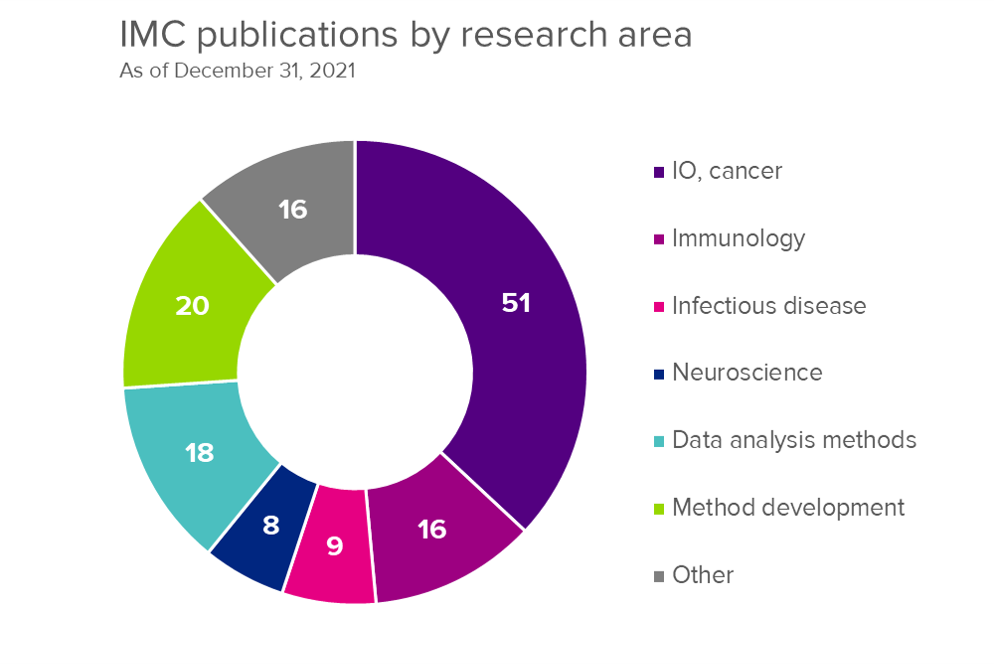
Immuno-oncology and cancer were the leading category with 37% of IMC publications in this research area. In addition to IMC technology being used in a wide range of applications, significant work has been done to develop methods and data analysis approaches.
For Research Use Only. Not for use in diagnostic procedures. Patent and License Information: www.standardbio.com/legal/notices. Trademarks: www.standardbio.com/legal/trademarks. Any other trademarks are the sole property of their respective owners. ©2025 Standard BioTools Inc. All rights reserved.