
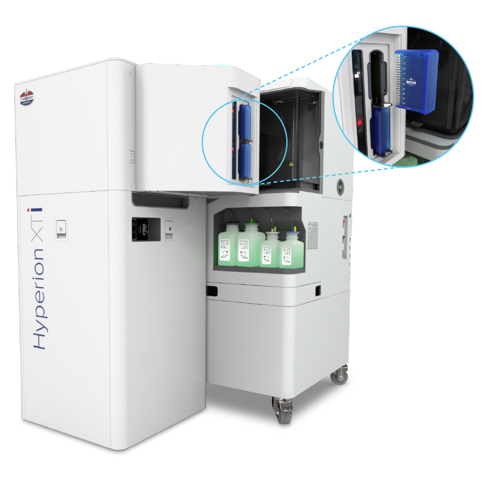
Hyperion XTi Imaging System
Unlocking new views in spatial biology
Introducing new groundbreaking capabilities for multiplexed tissue imaging at never-before-seen speeds and automation. The Hyperion™ XTi Imaging System is now available with three imaging modes for rapid and detailed analysis and automated slide loading for walk-away automation.
Discover the throughput and precision that is revolutionizing spatial biology.
See the new features
See the next generation in spatial biology
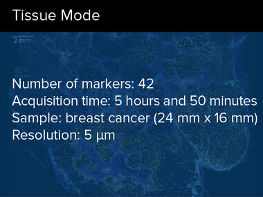
High-throughput multiplexed whole slide tissue imaging: The Hyperion XTi Imaging System now offers the ability to automate slide loading for continuous acquisition and new whole slide imaging modes for rapid and detailed analysis.
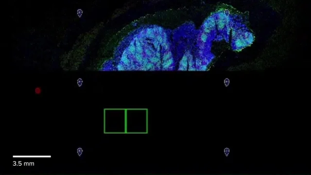
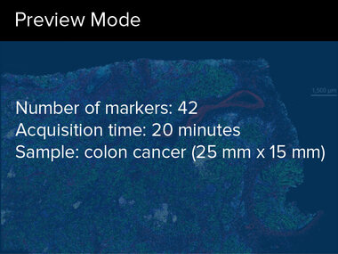
Visualize tissue heterogeneity in 20 minutes.

Take a deep dive into translational and clinical applications that explore new views of cellular composition within the tissue microenvironment.
TECHNOLOGY
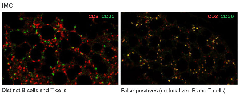
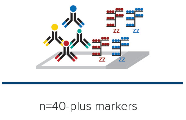
Powered by proven Imaging Mass Cytometry™ (IMC™) technology, the Hyperion XTi Imaging System is a multiplexed tissue imager that uses metal-tagged antibodies, instead of fluorophores, to simultaneously and reliably acquire 40-plus protein and RNA markers, without autofluorescence interference. IMC technology is the trusted choice for researchers and continues to be featured in high-impact publications – making it the most reliable tool for highly multiplexed imaging.
See data quickly.
Researchers can visualize 40-plus markers in real time, without managing time-consuming acquisition cycles – expediting the time to deeper analysis.
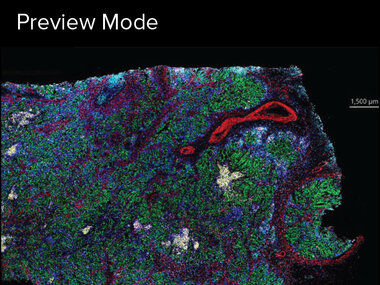
Represents Hyperion XTi Preview Mode for a large tissue section over 25 minutes. Researchers can select and view from any markers on the panel during acquisition.
See data clearly.
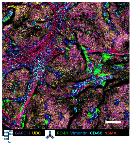
Clearly define areas in any tissue type ― including lung, bone marrow, colon and brain ― without background autofluorescence and spectral overlap.
Discover the power of Imaging Mass Cytometry technology
Image any tissue type with no autofluorescence interference
Co-detection of protein and RNA on the same tissue
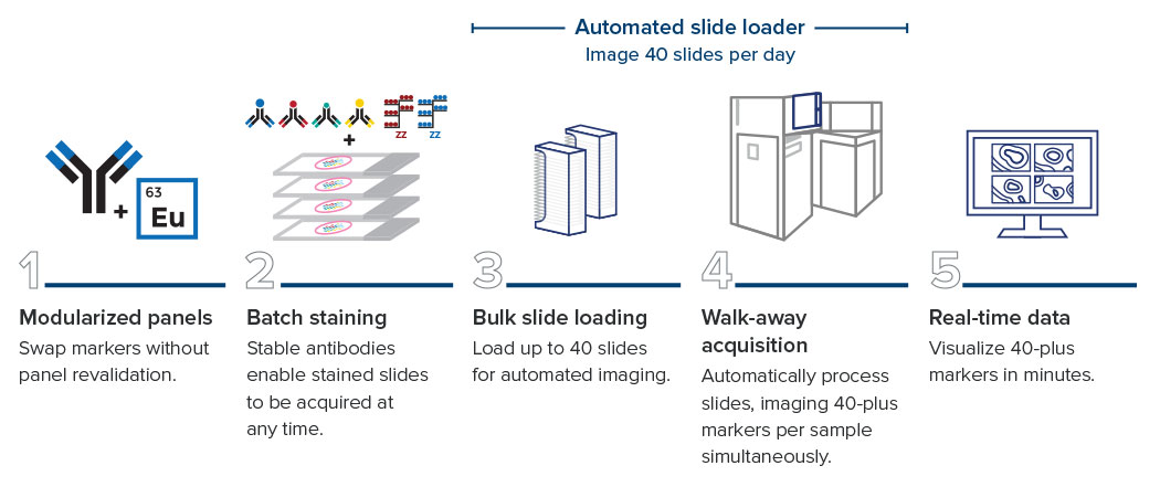
Automated slide loader for walk-away acquisition
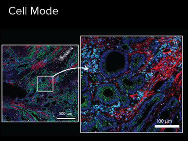
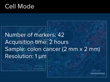
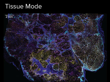
Three acquisition modes to accommodate any type of research
Batch staining workflow for high-volume studies
Multimodal capabilities with imaging and flow cytometry capabilities


Entering an era of new biology with rapid, high-throughput imaging
“Faster means more samples, more data. We can push through more of these small samples or we can do whole slides on the Hyperion XTi”
Hartland Jackson, PhD
Investigator
Lunenfeld-Tanenbaum Research Institute, Sinai Health
WORKFLOW
40 slides. 40-plus markers. 24 hours.
Load up to 40 slides and walk away.
Incorporate a new level of throughput and efficiency with the Hyperion XTi Slide Loader. The optional slide loader enables automatic loading and acquisition of up to 40 slides. It is equipped with a barcoded slide reader for easy sample identification, providing researchers with peace of mind when managing precious tissue samples.
See more from your samples.
Do more with your time.
Multiplexed imaging – without compromising speed
An end-to-end workflow to get results faster
A one-step staining and detection workflow
Imaging Mass Cytometry technology enables 40-plus markers that can be simultaneously stained, acquired and visualized. Other fluorescence-based approaches involve iterative rounds of staining, imaging and removal of fluorescent signals. The IMC workflow is without sequential immunostaining approaches, multiple slide treatment rounds or acquisition steps.
Protein and RNA co-detection
Detect protein and RNA on the same tissue sample to correlate transcriptional signatures and the spatial context of pathogens, host cells or protein sources. Explore this multi-omic workflow that combines RNAscope™ technology with IMC technology to visualize key RNA and protein markers in the same samples.
This image highlights RNA and protein co-detection in lung squamous cell carcinoma using the Hyperion XTi Imaging System and a 33-marker panel combined from three ready-to-go reagent kits.
Need to ship or store slides?
- All-at-once batch staining of all slides to reduce technical variation
- Acquire at any time from shipped and/or stored slides that have been stained
- Analyze previously banked tissue slides to be correlated to known clinical outcomes

Let us do the work for you.
Did you know Standard BioTools has an in-house service lab? Send us your samples and we will send you the data.
Applications
REAGENTS
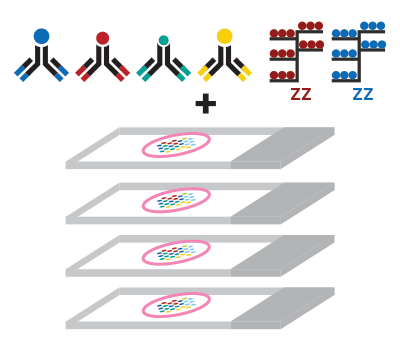
1. Start with ready-to-go human and mouse panels.
2. Easily add the Maxpar™ IMC Cell Segmentation Kit to your panel to solve the most important step in spatial biology.
3. Want to further customize your panel? Easily swap targets of interest from an ever-growing catalog of Maxpar IMC antibodies and panels.
View More PanelsHuman Immuno-Oncology IMC Panel, 31 Antibodies

The Human Immuno-Oncology IMC Panel, 31 Antibodies enables evaluation of critical pathophysiological parameters of the human tissue microenvironment using 31-plex Imaging Mass Cytometry™ (IMC™). This panel includes 31 rigorously tested and pathologist-verified antibodies that are also available in 6 modular subpanels for more flexible phenotyping options: Human Tissue Architecture IMC Panel, 4 Antibodies (201510), Human Stromal Cell IMC Panel, 4 Antibodies (201511), Human Basic Immune IMC Panel, 4 Antibodies (201518), Human Lymphoid IMC Panel, 4 Antibodies (201512), Human Myeloid IMC Panel, 6 Antibodies (201513), Human Cell Functional State IMC Panel, 5 Antibodies (201514), Human Epithelial and Mesenchymal IMC Panel, 4 Antibodies (201515).
Maxpar Neuro Phenotyping IMC Panel Kit

The Maxpar Neuro Phenotyping IMC™ Panel Kit is a seven-marker kit that includes validated human and mouse reactive antibodies against Iba1, GFAP, NeuN, S100β, MAP2, CD34 and Olig2. The markers serve to identify major cell types such as neurons, astrocytes, microglia, oligodendrocytes and endothelial cells and their functional state in the central nervous system (CNS). The kit is optimized for use in FFPE human and mouse tissue sections.
Human Cell Functional State IMC Panel, 5 Antibodies

The Human Cell Functional State IMC™ Panel, 5 Antibodies assesses the functional state of cells. This panel includes 5 pathologist-verified antibodies that enable assessment of cell proliferation, cytotoxic T cell activation, T cell exhaustion, and identification of regulatory T cells . This panel is optimized for use in FFPE human tissue sections.
Alzheimer's Disease IMC Panel, 3 Antibodies

The Alzheimer’s Disease IMC Panel is a bundle of three verified Maxpar OnDemand antibodies against Tau, phosphorylated Tau (pTau) and amyloid precursor protein (APP).
Maxpar® OnDemand™ Mouse Immuno-Oncology IMC™ Panel Kit

The Maxpar OnDemand Mouse Immuno-Oncology IMC Panel Kit (9100005) enables evaluation of critical pathophysiological parameters of the murine tumor microenvironment using 28-plex IMC. This panel kit includes 28 meticulously tested and verified antibodies and DNA markers. The kit consists of 4 modular subpanels: Mouse Tissue Architecture IMC Panel Kit (9100001), Mouse Cancer Cell Process IMC Panel Kit (9100002), Mouse Immune Phenotyping IMC Panel Kit (9100003), and Mouse Immune Activation IMC Panel Kit (9100004).
Poster: Novel Whole Slide Imaging Modes for Imaging Mass Cytometry Reveal Cellular and Structural Composition of Mouse Glioblastoma
Poster: Novel Whole Slide Imaging Modes for Imaging Mass Cytometry Unveil Extensive Cellular Heterogeneity in Human Gliomas
Poster: Next Generation of Spatial Biology: High-Thoroughput Multiplexed Imaging Mass Cytometry With Whole Slide Modes
Hartland Jackson, PhD

Entering an era of new biology
Daniel Schulz, PhD

Discovering a chemokine’s purpose in fighting tumors using RNA and protein co-detection
Yongpan Yan, PhD

An approach that combines molecular biology, immunology and cell biology tools to uniquely
Melissa Davis, PhD

Defining spatial characteristics using Imaging Mass Cytometry™ technology
Febe van Maldegem, PhD, and Karishma Valand

Imaging the dramatic remodeling of the lung tumor microenvironment
Denis Schapiro, PhD

The interactive histoCAT toolbox unifies the cytometry and imaging communities
David L. Rimm, MD, PhD

David Rimm on the value of Imaging Mass Cytometry™ for immunology applications at Yale
Corinne Ramos, PhD, MBA

Advancing drug development with molecular imaging
Bernd Bodenmiller, PhD

Bernd Bodenmiller on revealing new insights into tumors with Imaging Mass Cytometry™.
Akil Merchant, MD

Hear how researchers are using Imaging Mass Cytometry™ technology.
CyTOF Software v9.2.1
CyTOF™ Software v9.2.1 offers instrument control and data acquisition functionality for both imaging and flow applications with the Hyperion™ XTi Imaging System, CyTOF XT System and CyTOF XT PRO Sy...
MCD Viewer
MCD™ Viewer is post-acquisition data processing software that allows users to visualize, review and export Imaging Mass Cytometry™ data acquired with the Hyperion™ Imaging System and CyTOF™&nb...
Visiopharm Software
Phenoplex is a complete workflow software for multiplex tissue images in Spatial Biology, built on Visiopharm’s best-in-class AI, with interactive verification steps throughout.
ilastik
ilastik is an open source software for image analysis and machine learning. Using machine learning, ilastik software trains custom workflows for accurate cell segmentation and classification.
histoCAT
The histoCAT software is an innovative computational Imaging Mass Cytometry™ analysis toolbox that enables comprehensive analysis of cellular phenotypes and their interrelationships within the...
GET MORE INFORMATION
Interested in Standard BioTools™ Imaging Mass Cytometry products or need a quote? Contact our sales team for product orders, quotes or other inquiries.
For Research Use Only. Not for use in diagnostic procedures. Patent and License Information: www.standardbio.com/legal/notices. Trademarks: www.standardbio.com/legal/trademarks. Any other trademarks are the sole property of their respective owners. ©2025 Standard BioTools Inc. All rights reserved.