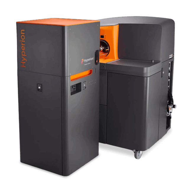
Hyperion Imaging System
Imaging Mass Cytometry: Visualize spatial biology. Confidently.
The Hyperion™ Imaging System allows access to single-cell insights. This includes a deeper understanding of the intricate spatial relationships of cells, pathways and phenotypes in the tissue microenvironment.

Technology
Imaging Mass Cytometry™ (IMC™) with the Hyperion Imaging System allows the generation of novel research hypotheses by surveying a high number of protein markers in one scan, saving precious sample, all while employing proven CyTOF® technology.
A precisely directed laser beam focused at 1 μm collects biological samples stained with metal-tagged Maxpar® antibodies from a region of interest. Next, discrete signals from each ionized metal tag are detected based on differences in mass instead of wavelength. Thus, IMC eliminates autofluorescence and achieves uniform staining with minimal background. Large panels can be rapidly designed with little concern about antibody order or label assignment.
When working with heterogeneous biology, assessing key spatial information is critical to driving a deeper understanding of both healthy and diseased tissue states. The Hyperion Imaging System is the ideal solution not only for this task, but to gain a better understanding of immunotherapy responses, with the intent to ultimately extend patient lives.

When speed matters, receive rapid answers for cohort analysis and clinical studies with the second-generation Hyperion+™ Imaging System.
Learn more
Workflow
1

Design
Design panels using pathologist-verified Maxpar antibodies conjugated to metal tags.
2

Stain
Stain tissues (FFPE or frozen) using familiar immunohistochemistry protocols.
3

Image
Image up to 40 protein markers in one scan at single-cell resolution using the Hyperion Imaging System.
4

Analyze
Analyze images in minutes using the MCD™ Viewer and easily export the resulting files for secondary analysis.
Publications
Keep current with how Imaging Mass Cytometry (IMC) is used to gain deep spatial insights – see articles, news, bibliographies, tips and more. Check out the IMC Trending Topics and Publications.
IMC TRENDING TOPICS & PUBLICATIONS
VIEW ALL PublicationsCyTOF Software
CyTOF® Software offers support for the imaging and cell suspension-based sample analysis to Hyperion™ Imaging System and Helios™ system users. The software has the built-in capability to switch sam...
CyTOF Software (Non-acquisition workstations only)
CyTOF® Software offers support for imaging analysis to Hyperion™ Imaging System users.
CyTOF Software v8.0
CyTOF® Software v8.0 offers support for cell suspension-based sample analysis to the CyTOF XT system. The software has the built-in capability for automated calibration, batch sample acquisition, a...
histoCAT
The histoCAT™ software is an innovative computational Imaging Mass Cytometry™ analysis toolbox that enables comprehensive analysis of cellular phenotypes and their interrelationships within the spa...
Indica Labs Software
The Indica Labs Highplex FL HALO® module combines ease of use and scalability with powerful analytics for streamlined biomarker analysis of highly multiplexed Imaging Mass Cytometry™ data. This mod...
MCD Viewer
MCD™ Viewer is post-acquisition data processing software that allows users to visualize, review, and export Imaging Mass Cytometry™ data acquired with the Hyperion™ Imaging System and CyTOF® Softwa...
Visiopharm Software
As high-plex IMC™ data continues to stretch our abilities to interrogate the tumor microenvironment, finding meaningful differences in your data becomes an ever more data-heavy and daunting challen...
Support
A critical success factor when adopting high-performance technology is acquiring the proficiency and support necessary to accelerate your research and discovery initiatives. Standard BioTools™ PRO Services are a comprehensive portfolio of solutions to help you maximize your investment, scale up productivity and empower your laboratory with greater access to professional resources.
Resources
Flyers & brochures
Site requirements
Application notes
Technical documents
User guides
Protocols
Specifications
The Hyperion Imaging System comprises a Hyperion Tissue Imager, a Helios system, and software for data acquisition and viewing.
Hyperion Tissue Imager dimensions*
Width
56 cm (22 in)
Height
134 cm (53 in
Depth
56 cm (22 in)
Weight
159 kg (350 lb)
Helios dimensions
Width
103 cm (41 in)
Height
132 cm (52 in)
Depth
87 cm (35 in)
Weight
320 kg (705 lb)
Chiller size
Width
38 cm (15 in)
Height
64 cm (25 in)
Depth
67 cm (27 in)
Weight
81 kg (178 lb)
Hyperion Imaging System
Channels
135
Mass range
75–209 amu
Abundance sensitivity
0.3% for 159Tb
Frequency
200 pixels/sec
Detection limit
≥400 copies per μm2
Dynamic range
4 orders of magnitude
Calibration
Automated
Operating system
Windows® 10 Enterprise
Data storage
OS: 240 GM and Storage: 8 TB
Switch time (between modes)
12 hr
Cross-cell contamination (redeposition)
≤2% (selected direction)
Crosstalk pixel to pixel
≤15% at 200 pixels/sec
Wet tissue thickness for full ablation
≤7 μm
Addressable sample size on slide
≥15 mm x 45 mm
Optical view of sample
≥250 μm x 250 μm
File type
TXT, multipage TIFF, OME-TIFF, MCD
Scan area
≥1 mm2/2 hr (@200 Hz)
Workstation
Operating system
Windows 10 Enterprise
Data storage
OS: 240 GB, Storage: 8 TB
CPU
Intel® Core i7
Memory
32 GB
Dimensions
Height 34.5 cm (14 in) , Width 13.4 cm (5 in), Depth 35.6 cm (14 in), Weight 8 kg (18 lb)
Monitor
86.3 cm (34 in)
Data file size**
MCD
4–5 MB/channel
TXT
6–8 MB/channel
* The Hyperion Tissue Imager must be placed directly in front of the Helios™
** 1,000 x 1,000 μm region of interest (ROI)
Unless explicitly and expressly stated otherwise, all products are provided for Research Use Only, not for use in diagnostic procedures. Find more information here.